Understanding “Saddle Nose” Risk in Traditional Surgery
If you’re researching septoplasty or rhinoplasty, you may run into the term “saddle nose.” In plain language, it describes a visible dip or collapse along the nasal bridge that can appear after surgery—and in some cases, it can also affect breathing. While this outcome is uncommon overall, surgeons understand why it happens and how to reduce the chances of it by protecting (or rebuilding) key support structures during surgery. (Rettinger 2008; Gadkaree et al. 2020) For a deeper explainer and visuals, see saddle nose deformity: https://www.clearpathnasal.com/blog/the-saddle-nose-deformity-what-you-need-to-know
Knowing the basics of saddle nose risk in traditional surgery helps you ask better questions before an operation and choose a plan that prioritizes long-term structure and airflow, not just short-term straightening.
What Is a “Saddle Nose” Deformity? (Simple Definition + What It Looks/Feels Like)
The structural issue: loss of support in the nasal dorsum (bridge)
A saddle nose deformity is a lowered or “collapsed” nasal bridge (the nasal dorsum) caused by weakened support. It can develop after trauma, but it’s also a known risk after prior nasal surgery—including septoplasty complications and certain rhinoplasty risks—when supporting cartilage is destabilized or removed. (Gadkaree et al. 2020)
A simple way to picture it: if the bridge of the nose is like a gently arched beam, the septum and surrounding cartilages help hold that beam up. When the support is weakened, the “beam” can settle—creating a dip that may be subtle at first and more noticeable as swelling resolves.

Why it’s not just cosmetic—breathing can be affected
The nasal bridge area isn’t only about appearance. The middle portion of the nose helps maintain the shape of the airway. When that support weakens, the middle vault can narrow the internal nasal valve (a key airflow bottleneck), leading to obstruction even if the septum was technically straightened. Patients may notice a new “pinched” feeling or collapse when breathing in. (Gadkaree et al. 2020; Rettinger 2008) Learn more about the internal nasal valve and why it matters for breathing: https://www.clearpathnasal.com/blog/understanding-the-nasal-valve-why-it-matters-for-breathing
A patient-friendly example: someone may say, “My nose looks a little flatter than before—and I feel more blocked when I take a deep breath,” especially during exercise or when allergies flare.
• Bottom line: saddle nose reflects lost structural support, which can also narrow key airflow zones. •

How Traditional Septoplasty or Rhinoplasty Can Increase Saddle Nose Risk
It’s not only “over-resecting the hump”
A common misconception is that saddle nose only happens when too much bridge/hump is removed during cosmetic rhinoplasty. In reality, risk can also rise when the septal support system is weakened, fractured, or over-resected—especially in procedures focused on function, like septoplasty. (Rettinger 2008; Gadkaree et al. 2020) For a broader context of procedural risks, see septoplasty complications: https://www.clearpathnasal.com/blog/septoplasty-complications-how-clearpath-minimizes-risks
A particularly important area is the caudal septum support (the front/bottom portion of the septal cartilage). If this region is destabilized without adequate stabilization or reconstruction, the connection between the septum, bridge, and tip can be compromised.
The key anatomy in plain English (why the septum is a “keystone”)
Think of the septum as a central support pillar inside the nose. It helps hold up:
- the upper lateral cartilages (middle vault/bridge region)
- the alignment and strength of the nasal dorsum
- portions of tip support
When that pillar is weakened, the bridge can settle downward and the internal nasal valve region can narrow—linking cosmetic changes with breathing symptoms. (Gadkaree et al. 2020)
A quick analogy: the “L-strut” as the nose’s frame
Surgeons often talk about preserving a supportive “L-shaped” portion of cartilage (the L-strut) during septal work. If you imagine the nose as a small tent, the L-strut functions like a key tent pole: remove too much or leave it unstable, and the structure above it can sag. That’s why prevention is less about “making things straight” and more about keeping (or rebuilding) a stable framework.
• Support preservation—not just straightening—is what lowers risk. •

How Common Is Saddle Nose After Septoplasty? (What Studies Report)
Reported rates vary depending on definition and detection
Published reports vary because studies define “noticeable change” differently and follow patients for different lengths of time. Earlier rhinoplasty risk and septoplasty literature has cited noticeable cosmetic change after septoplasty in the range of ~0.4%–3.4%. (Rettinger 2008)
It’s also worth remembering that early swelling can temporarily mask contour changes. A result that looks “fine” in the first few weeks may look different months later—one reason careful follow-up matters.
A specific data point patients can understand
In one retrospective series of septoplasty patients, intraoperative saddle nose was identified in 108 of 2,106 patients (5.1%). (Lee et al. 2019)
“Intraoperative” means the concern was recognized during the operation—it does not automatically translate to the long-term rate of visible saddle nose after healing. To avoid confusion: this statistic reflects what surgeons saw and managed in real time, not the final cosmetic outcome months later. Still, it’s a useful reminder that structural weakening can happen if support is compromised, and that surgeons may need to pivot to stabilization techniques when they see risk emerging.
• Rates are low overall, and consistent follow-up helps catch changes that emerge as swelling resolves. •
Who Is at Higher Risk? (Patient-Friendly Risk Factors)
No one can predict risk perfectly, but some factors make surgeons more cautious about preserving or rebuilding structure:
Severe caudal septal deviation
When deviation is severe—especially in the caudal septum—more manipulation may be needed to reposition cartilage. Without careful planning, that can increase the chance of destabilizing the septal L-strut (the critical supportive framework). (Lee et al. 2019)
A practical example is a septum that bends strongly near the nostril entrance (not just deeper inside). Correcting that area can be mechanically demanding, which is why technique and stabilization choices matter.
Intraoperative septal-cartilage fracture
An unintended cartilage fracture can weaken support significantly, increasing the odds of dorsum settling or middle vault collapse. (Lee et al. 2019)
Clinicians sometimes describe this as a “hinge” or “buckling” moment: once cartilage loses integrity, it may not hold shape during healing unless it’s reinforced.
Female sex (as reported in one study)
One series reported female sex as a significant risk factor. The underlying reasons aren’t fully established and may relate to anatomy or cartilage characteristics, but it’s best viewed as a study finding, not a personal prediction. (Lee et al. 2019)
• Your anatomy and the intraoperative course shape risk more than any single demographic factor. •
Prevention: What Surgeons Do to Reduce the Risk (And What Patients Can Ask About)
Prevention centers on one principle: support first. Many septoplasty complications (including saddle changes) become less likely when the plan protects or reconstructs structural stability. (Gadkaree et al. 2020; Rettinger 2008)
Preserve caudal septal support and avoid overly aggressive cartilage removal
Surgeons typically aim to preserve the key supporting struts of cartilage rather than removing too much. This is especially important when addressing deviations that involve the caudal septum. (Gadkaree et al. 2020; Rettinger 2008) Some specific techniques are designed to prevent saddle deformity by reinforcing the caudal segment and L‑strut during septoplasty. (Raeessi et al. 2008)
In patient terms: you want a plan that fixes the blockage without “taking the beams out of the house.” Straightening is helpful only if the structure remains strong afterward.
Stabilize mobile or dislocated cartilage during surgery
Depending on the anatomy and goals, surgeons may use structural techniques such as:
- structural septal struts (to reinforce support)
- spreader grafts (to support the middle vault and help protect internal valve function)
- temporary stabilization sutures (to prevent shifting during healing)
These choices are individualized, but the theme is consistent: maintain dorsal height and protect airflow pathways. (Gadkaree et al. 2020; Rettinger 2008)
Patient question checklist (quick bullets)
Consider asking:
- How will you preserve the L-strut and caudal septum support?
- If cartilage is weakened, what stabilization or grafting options do you use?
- How do you evaluate and protect the internal nasal valve to prevent collapse?
For broader context, see septoplasty complications: https://www.clearpathnasal.com/blog/septoplasty-complications-how-clearpath-minimizes-risks
• Ask how your surgical plan keeps or rebuilds support while improving airflow. •

Signs Something Isn’t Right After Surgery (When to Call Your Surgeon)
Appearance changes to watch for
- A new dip along the bridge
- Loss of dorsal height you didn’t expect
- New asymmetry that doesn’t improve as swelling goes down
Function changes to watch for
- Worsening nasal blockage, especially higher in the nose (middle vault area)
- A “pinched” sensation
- New collapse when you inhale (which may suggest internal nasal valve collapse) (Gadkaree et al. 2020)
Timing
Early healing involves swelling and shifting sensations. The key is persistence: if a change continues beyond the expected swelling phase—or if breathing worsens rather than improves—follow up with your surgeon rather than trying to self-diagnose. A useful way to frame it is, “Is this getting steadily better week by week?” If not, that’s a reasonable reason to check in.
• If changes persist or breathing worsens, contact your surgeon rather than waiting it out. •
If Saddle Nose Happens: Treatment Options by Severity
Limited defects: cartilage grafting
Smaller depressions may be corrected with cartilage grafting using septal cartilage (if available) or conchal (ear) cartilage. (Gadkaree et al. 2020) Think of this like resurfacing a low spot: the goal is to restore smooth contour while supporting the area so it doesn’t collapse again.
More extensive collapse: structural reconstruction
When support loss is larger, reconstruction may require stronger structural material—often rib cartilage graft (costal cartilage) or bone grafts—to rebuild dorsal height and maintain airway stability. (Gadkaree et al. 2020; Riechelmann et al. 2004) This is closer to rebuilding the underlying frame, not just filling a dip, because the middle vault and valve region may need durable support.
Goal is both form + function
Successful reconstruction should address:
- the visible bridge contour (dorsal height and smoothness), and
- the airway—especially the internal valve region—so that breathing improves, not just appearance. (Gadkaree et al. 2020)
• Reconstruction aims to restore both contour and airflow, tailored to severity. •

Minimally Invasive Approaches and the “Support Preservation” Conversation (ClearPath Context)
Why less dissection may matter for structural stability
Because saddle nose risk in traditional surgery is fundamentally tied to structural destabilization, many patients ask whether less-disruptive approaches could help preserve support. In general, techniques designed to reduce dissection and tissue disruption may reduce certain morbidities—but outcomes still depend heavily on anatomy, indications, and surgeon technique.
In traditional septoplasty, lifting mucoperichondrial flaps and manipulating cartilage is sometimes necessary, but more dissection can also mean more tissue trauma and destabilization risk in certain cases. Approaches that aim to reduce flap elevation may, in theory, help reduce specific dissection-related issues (not a guarantee, and not appropriate for every deviation pattern).
ClearPath Nasal Balloon (CNB): design intent (high-level)
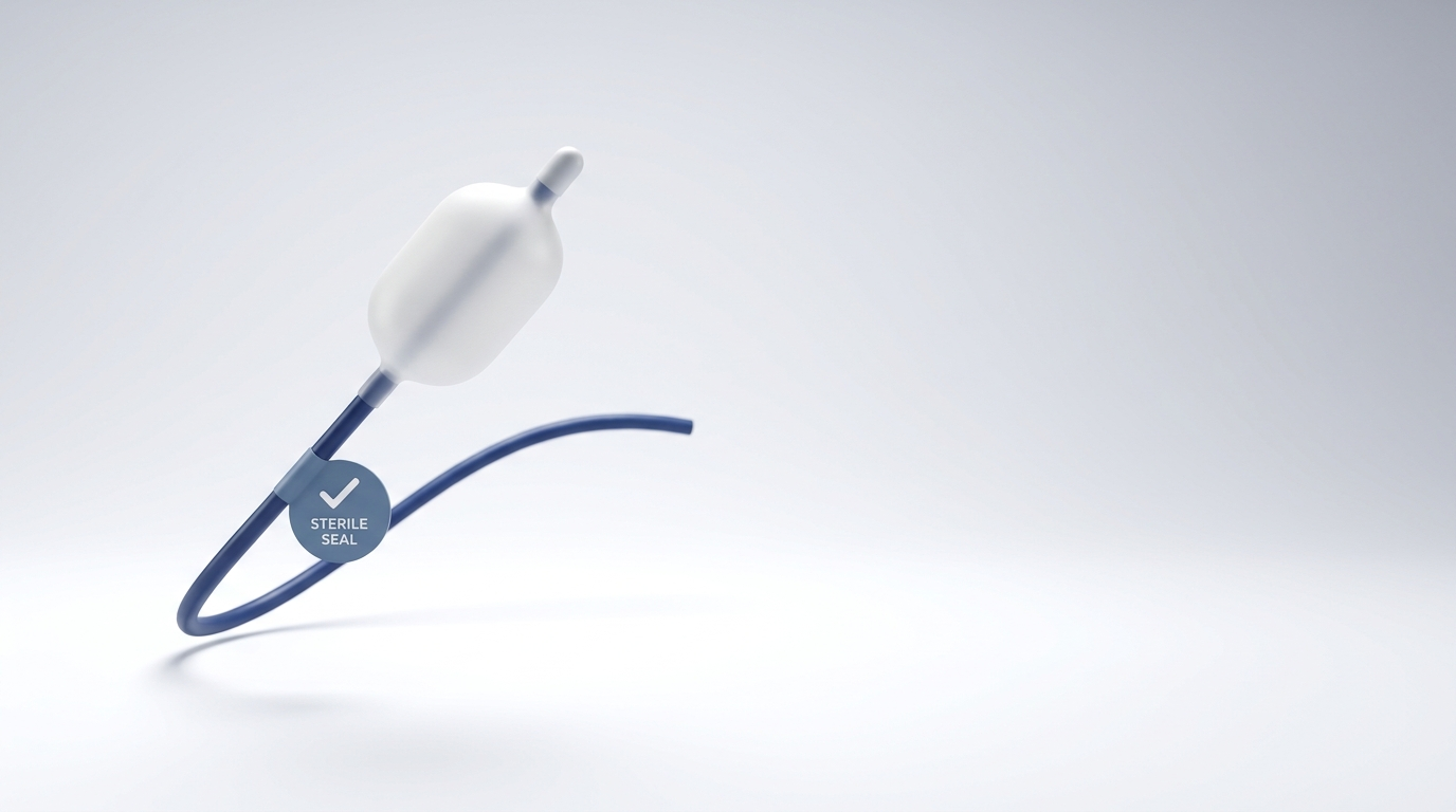
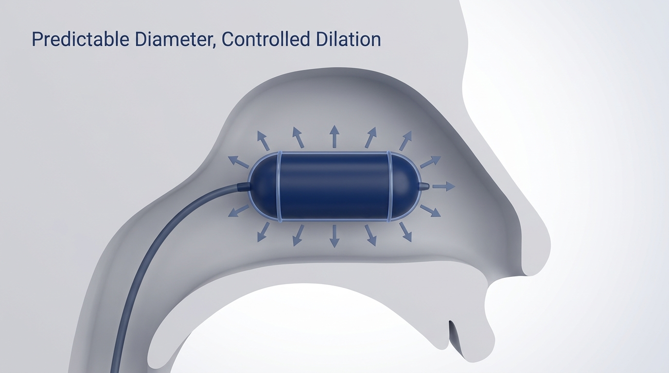
ClearPath Nasal Balloon (CNB) is a device used in a minimally invasive, endoscopic approach intended to mobilize certain septal deviations. It uses a force-directed balloon and shaped spatula to mobilize bony deviations while aiming to reduce or avoid mucoperichondrial flap elevation, packing, and prolonged anesthesia compared with traditional septoplasty.
Safety and early outcomes (patient-friendly summary)
In an early single-center retrospective series of 107 consecutive cases, no major complications were reported (including no septal perforation, hematoma, synechia, or conversion to traditional flap-based surgery). These data are preliminary and retrospective; larger prospective studies are still needed.
Imaging + symptom improvement (simple explanation)
In a small CT-imaging subgroup (n=23), septal alignment improved on average 45% at maximal deviation and 22% at the nasolacrimal duct level (p=0.005). In a SNOT-22 subgroup (n=43 at 1 month), symptom scores improved by 23.8% on average (58.7 → 44.8; p=0.02). Because the data are retrospective and the CT subgroup is modest, larger prospective studies are needed to confirm durability and generalizability. For more context on differences in approach and recovery, see ClearPath vs traditional septoplasty: https://www.clearpathnasal.com/blog/clearpath-vs-traditional-septoplasty-recovery-safety-results
• Less dissection can reduce certain morbidities, but suitability and outcomes depend on your anatomy and the surgeon’s assessment. •
Conclusion: How to Make a Lower-Risk Surgical Plan
The big idea is simple: a saddle deformity is fundamentally a support problem. Saddle nose risk in traditional surgery tends to rise when septal support—especially caudal septum support—is destabilized, fractured, or over-resected, sometimes leading to middle vault weakness and internal nasal valve collapse. (Lee et al. 2019; Gadkaree et al. 2020; Rettinger 2008)
If you’re planning septoplasty or considering rhinoplasty, choose a surgeon who can clearly explain:
- how they preserve or rebuild the septal L-strut,
- how they protect nasal valve function, and
- what they’ll do if cartilage is weak (including stabilization or grafting plans).
If you’re weighing options, ask your local ENT surgeon whether you’re a candidate for a minimally invasive alternative—such as an approach using the ClearPath Nasal Balloon—and how each option affects long-term structural support and nasal-valve airflow.
• Choose a plan that prioritizes support and airflow first, not just immediate straightness. •
References
- Gadkaree SK, et al. Review of Literature of Saddle Nose Deformity Reconstruction. 2020. (PMC7752089)
- Lee J, et al. Risk Factors for Intraoperative Saddle Nose Deformity in Septoplasty Patients. 2019. (PubMed 30937560)
- Rettinger G. Risks and Complications in Rhinoplasty. 2008. (PMC3199839)
- Riechelmann H, et al. Three-Step Reconstruction of Complex Saddle Nose Deformities. 2004. (JAMA Otolaryngology)
- Raeessi M, et al. A New Technique During Septoplasty to Prevent Saddle Nose. 2008. (Wiley)
- Dillard J, et al. Rhinology Online. Preliminary single-center retrospective series; publication timing and peer-reviewed status should be verified; larger prospective studies are needed.
This article is for educational purposes only and is not medical advice. Please consult a qualified healthcare provider for diagnosis and treatment.

Dr. David G. Dillard
Author
Share it with others!