Synechia Formation: A Risk of Traditional Surgery
Scar tissue is a normal part of healing—but sometimes healing goes a step too far. After surgery or inflammation, tissues that should remain separate can “stick” together. That sticking can slow recovery, distort anatomy, or create new symptoms that weren’t part of the original problem.
This article explains synechia formation in plain English, why it’s more likely after more disruptive (“traditional”) surgery, what symptoms to watch for, and how modern, tissue-preserving approaches—including the ClearPath device in nasal surgery—aim to reduce complication burden.
What “Synechia Formation” Means (Plain-English Definition)
Synechiae (pronounced “sin-NECK-ee-eye”) are scar-like adhesions. In other words: two tissues that shouldn’t be attached end up sticking together after inflammation, trauma, or surgery. [1]
A useful analogy: think of two moist surfaces healing like two pages of a wet book. If they’re pressed together while drying, they can fuse. In the body, that “fusing” is scar tissue forming an unintended bridge.
It’s understandable to feel anxious when you hear unfamiliar medical terms. The key thing to know is that synechiae are a risk—not an inevitability—and they’re often manageable, especially when detected early.

Synechiae vs. “Adhesions” vs. “Scar Tissue”
- Scar tissue is the body’s repair material.
- Adhesions describe scar tissue that creates an abnormal “bridge” between surfaces.
- Synechiae are a specific type of adhesion doctors commonly describe in areas where delicate structures sit close together (like inside the eye or inside the nose).
Surgeons care about adhesions because they can restrict normal movement, block flow, or make healing more complicated—sometimes in ways that don’t show up until weeks after surgery.
Where Synechiae Can Occur (Different Specialties, Same Problem)
- Ophthalmology (eye care): Synechiae can form between structures such as the iris and nearby tissues. Depending on location and severity, they can affect eye pressure and visual outcomes. [1]
- ENT (inside the nose): After nasal surgery, internal nasal surfaces can heal together unintentionally. When that happens, patients may notice persistent blockage or reduced airflow—sometimes on the same side the surgery was meant to open.
In short: synechiae are unintended tissue bridges that can form during healing—and they’re often manageable when recognized early.

Why Traditional Surgery Can Increase Synechia Risk
A helpful rule of thumb is: more tissue disruption can mean more postoperative inflammation, which can raise adhesion risk. That said, risk is multifactorial and varies by patient, anatomy, and procedure type. If you’re exploring nasal procedures, you may also want to read about septoplasty risks and complications: https://www.clearpathnasal.com/blog/septoplasty-complications-how-clearpath-minimizes-risks
Inflammation Is the Main Driver
In eye surgery, synechiae are strongly linked with postoperative inflammation, and the risk is higher in people with inflammatory eye disease (such as uveitis). [2] This relationship—between inflammation and adhesions—shows up across many areas of medicine: more inflammation generally means more “repair signaling,” and more repair signaling can mean more scar formation. Clinicians often summarize it simply: the calmer the tissue after surgery, the lower the chances it heals in an overly sticky way. Still, calmer tissue is a goal—not a guarantee.
Longer or More Complex Operations Raise Risk
Complexity matters. In combined retinal/cataract procedures (phacovitrectomy), studies report widely varying synechiae rates—from 3.7% to 30.8%—and one meta-analysis found posterior synechiae in about 18% of patients. [3] Risk rises with factors that often reflect a more complicated case, such as longer duration or additional surgical steps (for example, silicone-oil tamponade). [3] These ophthalmology figures illustrate the principle but don’t predict rates for other specialties; actual risks are procedure- and anatomy-specific.
Less disruption often means less inflammatory burden—but individual risk depends on the specific operation, your underlying condition, and how your body heals.

Why Synechiae Matter Clinically (What They Can Disrupt)
Adhesions aren’t just a “paperwork diagnosis.” Depending on where they form, synechiae can:
- Block flow (like fluid pathways)
- Pull structures out of position
- Reduce function (airflow, movement, or drainage)
- Slow recovery and increase the chance of needing additional treatment
Even when the adhesion is small, its location can make it clinically meaningful—especially in tight spaces (like the eye’s drainage angle or narrow nasal passages).
Examples From Eye Surgery (Well-Documented Consequences)
- After glaucoma surgery: Peripheral anterior synechiae can obstruct the surgical opening after trabeculectomy, contributing to eye pressure elevation and reduced surgical success. [4]
- After cataract surgery in angle-closure disease: Pre-existing synechiae can limit how much the front chamber of the eye deepens, and they may be associated with larger refractive errors after cataract surgery. [5]
- In severe situations: Synechiae can contribute to glaucoma and permanent vision loss. [1]
A practical way to picture this: if normal anatomy is like a doorway, synechiae can act like a doorstop placed in the wrong spot—reducing the opening or changing how fluid and structures move.
Small adhesions in tight spaces can have outsized effects on function and recovery.
Who Is at Higher Risk for Synechia Formation?
While anyone can develop adhesions, risk increases when inflammation is higher, anatomy is tighter, or the procedure is more complex.
Medical & Anatomic Risk Factors
- Higher inflammatory tendency (for example, uveitis in eye care) is associated with more postoperative complications tied to inflammation. [2]
- Pre-existing adhesions: For instance, peripheral anterior synechiae in angle-closure eyes can influence outcomes and recovery patterns after cataract surgery. [5]
If you’ve been told you already have adhesions, that doesn’t automatically mean you’ll get more—just that your surgeon may plan closer surveillance and more proactive inflammation control.
Procedure-Related Risk Factors (Complexity, Duration, Tamponade)
In a 2024 study of phacovitrectomy, risk factors associated with posterior synechiae included retinal detachment surgery, silicone-oil tamponade, and procedures lasting more than 93 minutes. [3] These details help explain why two people can have “the same surgery” yet different risk profiles: the underlying diagnosis and the steps required can change the inflammatory load.
Knowing your personal risk helps your care team tailor prevention, follow-up, and treatment plans to you.
Signs and Symptoms Patients Might Notice After Surgery
Symptoms depend on the body area involved. In general, contact your surgical team if you notice:
- Recovery that feels stalled or delayed
- Symptoms that improve then worsen
- A new sense of blockage, tightness, or pulling
- Unexpected changes in function (such as new blur, pressure sensations, or worsening nasal airflow)
In ENT recovery specifically, a patient might describe it as: “I expected congestion early, but this feels like one side never really opens up.” In eye care, it might be a sense of worsening blur, pressure, or slower-than-expected visual recovery—always worth checking.
When to Call the Surgeon Urgently
Seek urgent guidance for sudden worsening, severe pain, rapidly decreasing function, heavy bleeding, or any symptom your post-op instructions flag as urgent.
If recovery stalls or reverses—especially after an initial improvement—get in touch promptly.

How Clinicians Detect and Monitor Synechiae
Postoperative Exams Matter
Follow-ups aren’t just routine—they’re timed to catch issues early, when treatment is often easier. Early synechiae can be subtle, and prompt identification may reduce the chance that an adhesion becomes firm or restrictive. One reason surgeons emphasize “keep the appointment even if you feel fine” is that adhesions can be visible on exam before they’re obvious in day-to-day symptoms.
Imaging/Visualization (Specialty-Specific)
- Eye care: slit-lamp exam and gonioscopy help clinicians see fine internal adhesions and evaluate drainage angles.
- ENT: nasal endoscopy allows direct visualization inside the nose to check healing surfaces and airflow pathways.
Early checks can reveal quiet problems before they harden into lasting issues.
Prevention: How to Lower the Risk of Synechia Formation
Control Inflammation Early and Consistently
Because inflammation is such a key driver, postoperative anti-inflammatory plans matter. In uveitis and cataract contexts, controlling inflammation is a major strategy to reduce complications tied to healing responses. [2] From a patient standpoint, this is where adherence pays off: taking drops/sprays exactly as directed and not stopping early can meaningfully support smoother healing.

Surgical Approach Matters: Less Trauma Can Mean Less Scarring
Across specialties, a tissue-preserving plan—less disruption, efficient technique, fewer “extra” steps when safe—may reduce downstream scarring risk. That concept aligns with findings that complexity and longer operative time are associated with higher synechiae risk in certain settings. [3] For readers comparing nasal approaches, this background pairs well with reducing flap elevation: https://www.clearpathnasal.com/blog/reducing-flap-elevation-with-clearpath
Diligent inflammation control and gentle technique work together to lower the odds of overly “sticky” healing.
Management Options If Synechiae Do Form
Watchful Waiting vs. Intervention
Not every adhesion needs treatment. Many are small and cause minimal symptoms. Management depends on:
- What the adhesion is doing (blocking, pulling, narrowing)
- How the patient feels and functions
- Whether it threatens long-term outcomes
In other words: the goal isn’t to “treat a finding,” but to protect function and reduce downstream complications.
Adhesion Release: Laser-Based vs. Traditional Revision
In eye care, argon laser synechiolysis has been reported as a way to reverse certain adhesions after trabeculectomy—potentially avoiding some risks of returning to the operating room for surgical revision. [4]
Minimally Invasive Options (Where Appropriate)
In some fields, minimally invasive approaches have been developed to reduce complication burden compared with more traditional incisional surgery, while recognizing that effectiveness and candidacy vary by condition and anatomy. [6] For example, microinvasive glaucoma surgery (MIGS) is generally described as having a lower serious-complication profile than traditional incisional glaucoma surgery, although its pressure-lowering effect can be less in some situations. [6]
Treat the impact—not just the appearance—of an adhesion, using the least disruptive option that addresses the problem.
What “Minimally Invasive” Can Look Like (Example From Nasal Surgery Safety Data)
Minimally invasive design aims to reduce dissection-related morbidity—often by limiting tissue lifting, reducing trauma, and supporting smoother recovery. In nasal surgery, a modern example is balloon-assisted septoplasty using the ClearPath device. For a broader comparison, see ClearPath vs. traditional septoplasty: https://www.clearpathnasal.com/blog/clearpath-vs-traditional-septoplasty-recovery-safety-results
The goal is the same across techniques: achieve the intended outcome with less tissue stress when appropriate.
Balloon-Assisted, Tissue-Preserving Intent (ClearPath CNB Example)
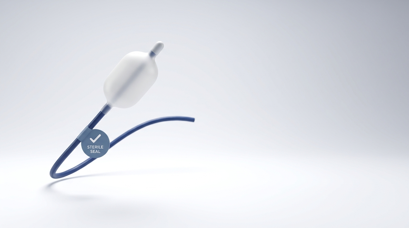
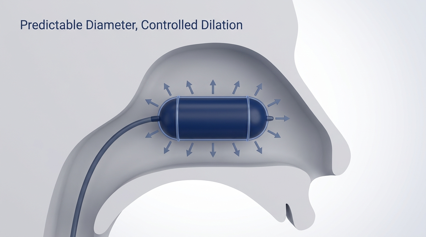
The ClearPath Nasal Balloon (CNB) technique uses a force-directed balloon and a shaped spatula to mobilize septal deviations while aiming to reduce or avoid mucoperichondrial flap elevation, packing, and prolonged anesthesia compared with traditional septoplasty approaches. [7] If you want a step-by-step explainer, see what is balloon-assisted septoplasty?: https://www.clearpathnasal.com/blog/what-is-balloon-assisted-septoplasty
Safety Snapshot (CNB Septoplasty Series)
In a single series of 107 consecutive CNB septoplasty cases, investigators reported no major complications, including no septal perforation, hematoma, synechia, or conversion to traditional flap-based surgery. [7] This is an encouraging early experience but not comparative, randomized evidence; results can vary by patient factors, surgeon, and setting.
Early series can guide expectations, but broader, controlled studies are needed to confirm safety and effectiveness.
Outcomes Patients Care About (Quality of Life + Imaging)
- In a CT subgroup (n=23), mean septal alignment improved 45% at the point of maximal deviation and 22% at the nasolacrimal duct (both p=0.005). [7]
- In 43 patients with one-month surveys, SNOT-22 scores improved from 58.7 to 44.8 (about 23.8% improvement, p=0.02). [7]
These outcomes reflect the broader goal: reduce symptoms while supporting safer healing—potentially lowering conditions that can lead to synechia formation in the first place. As always, confirm whether a given approach fits your anatomy and goals.
Patient-centered outcomes are promising, but durability and generalizability should be validated in larger cohorts.
Patient Checklist: How You Can Support Healing After Surgery
Do’s
- Take anti-inflammatory medications exactly as prescribed
- Keep every follow-up appointment (even if you feel “fine”)
- Report new or worsening symptoms early—don’t wait for the next scheduled visit
- For practical pointers, see septoplasty recovery tips: https://www.clearpathnasal.com/blog/septoplasty-recovery-tips-getting-back-to-normal-faster
Don’ts
- Don’t stop drops, sprays, or medications abruptly without guidance
- Don’t ignore a recovery that isn’t progressing as expected
Small, consistent steps—medication adherence, early reporting, and kept appointments—support calmer healing.
FAQ (Patient-Focused)
What causes synechia formation after surgery?
Most often, postoperative inflammation leads tissues to heal together when they’re close in proximity. Greater tissue disruption or longer, more complex operations can increase risk, but overall risk is multifactorial. [2][3]
Are synechiae permanent?
They can be, especially if they mature and become fibrotic. But some synechiae are minor, and others can be treated—particularly when caught early.
How are synechiae treated—does it always require another surgery?
Not always. Some adhesions are monitored. Others may be released with targeted procedures; in eye care, laser synechiolysis is one documented option in select cases. [4]
Does minimally invasive surgery lower the risk of synechiae?
Minimally invasive techniques are designed to reduce tissue disruption and inflammation—two factors linked to adhesion risk. Evidence and outcomes vary by specialty, procedure, and patient anatomy. In nasal surgery, ClearPath CNB septoplasty has reported a favorable safety profile in a single 107-case series (non-comparative), including no synechiae observed. [7]
When should I worry about symptoms after surgery?
Call your surgeon if symptoms worsen suddenly, pain becomes severe, function declines rapidly, or recovery stalls—especially after an initial period of improvement.
Conclusion: The Practical Message
Synechia formation is a known risk of healing after surgery—particularly when inflammation is high or the procedure is long and complex. [2][3] The best protection is a combination of strong inflammation control, careful postoperative monitoring, and timely treatment when an adhesion becomes clinically important (including synechiolysis options in certain settings). [4] When appropriate, less disruptive approaches may help reduce complications linked to tissue trauma and postoperative inflammation. In ENT, balloon-assisted septoplasty using the ClearPath device is one example of a tissue-preserving alternative to traditional procedures, with encouraging early safety and outcomes data in a single-center consecutive series. [7] If you’re considering septal surgery, ask your local ENT whether ClearPath balloon-assisted septoplasty is an option for your anatomy and goals.
The practical bottom line: steady inflammation control, early follow-up, and right-sized interventions help keep healing on track.
References
[1]: Cleveland Clinic. Synechiae (Eye). https://my.clevelandclinic.org/health/diseases/24874-synechiae-eye
[2]: Al‑Essa RS, et al. New insights into cataract surgery in patients with uveitis. 2022. https://pmc.ncbi.nlm.nih.gov/articles/PMC9535913/
[3]: Pesoa S, et al. Posterior synechia formation after phacovitrectomy—Predicting factors and the role of short-acting mydriatics. Acta Ophthalmologica. 2024. https://pmc.ncbi.nlm.nih.gov/articles/PMC11149058/ and https://onlinelibrary.wiley.com/doi/10.1111/aos.15760
[4]: Kaur M, et al. Reversal of Peripheral Anterior Synechiae After Trabeculectomy With Argon Laser Synechiolysis. 2024.
[5]: Lee et al. The effects of peripheral anterior synechiae on refractive outcomes after cataract surgery in eyes with primary angle-closure disease. 2021. https://pmc.ncbi.nlm.nih.gov/articles/PMC8036052/
[6]: EyeWiki. Microinvasive Glaucoma Surgery (MIGS). https://eyewiki.org/Microinvasive_Glaucoma_Surgery_(MIGS)
[7]: Dillard J, et al. Rhinology Online. 2026;9:18–23. doi:10.4193/RHINOL/25.018
This article is for educational purposes only and is not medical advice. Please consult a qualified healthcare provider for diagnosis and treatment.

Dr. David G. Dillard
Author
Share it with others!