Septoplasty for Spurs: When Do You Need a Flap?
If you’ve been told you have a septal spur, it’s normal to wonder: “Do I actually need surgery?” And if you’re considering septoplasty, you may also hear your ENT mention a “flap”—which can sound intimidating if no one explains it in plain language.
This guide breaks down what septal spurs are, when septoplasty may help, what a mucoperichondrial/mucoperiosteal flap is, and why protecting the septum’s lining is a big part of reducing septal perforation risk. We’ll also explain where endoscopic septoplasty and balloon-assisted approaches fit into the conversation—starting with a key limitation: balloon tools, including ClearPath, are generally not designed to shave down or remove a true bony/cartilaginous spur, so traditional instruments are typically required for spur work.
A useful theme to keep in mind: septoplasty is generally aimed at a symptomatic septal deformity (most commonly nasal obstruction), not an incidental imaging finding. (StatPearls—Septoplasty, NCBI Bookshelf)
What Is a Septal Spur (and Why It Matters)?
A septal spur is a sharp ridge of cartilage and/or bone along the nasal septum (the “wall” between your nostrils). Think of it like a small bump or ledge sticking out from that wall. Some people never notice it; others feel it because of where it sits and what it touches.
Many spurs are found incidentally and do not automatically need treatment. Septoplasty decisions usually start with symptoms—most often nasal blockage—and then ask whether the exam findings plausibly explain those symptoms. (StatPearls)
A quick, concrete example
- Person A: breathes well day and night; spur is an incidental note → surgery often isn’t needed.
- Person B: consistently feels “stuck” on one side, and endoscopy shows the spur narrowing airflow or contacting nearby tissue → septoplasty may be worth discussing.
Septal spur vs. deviated septum (what’s the difference?)
A deviated septum is often a broader curve or shift of the septum to one side. A spur is typically more focused and sharper—like a pointed ridge. You can have one without the other, but they commonly coexist.
If you’d like a deeper anatomy overview, see our post on septal spurs and bony deformities: https://www.clearpathnasal.com/blog/understanding-septal-spurs-and-bone-deformities.
Common symptoms linked to a spur
- Nasal obstruction (often worse on one side)
- Recurrent nosebleeds in selected cases (airflow turbulence can dry/irritate the lining, but epistaxis has many other causes)
- Possible “contact point” irritation (when a spur touches a turbinate); the link with facial pain/headache is uncertain and remains controversial, and other causes should be ruled out first
(StatPearls; AAO-HNS Clinical Consensus Statement, 2015)
In short: imaging can find spurs in people who breathe well—symptoms plus exam are what count most.

When Does a Septal Spur Actually Need Septoplasty?
“A spur on CT” isn’t the same as “a spur causing symptoms.” It’s very common for imaging to show a spur even in people who breathe fine. The key question is whether the anatomy matches your symptoms and what your ENT sees on exam.
In other words, septoplasty for a septal spur is typically considered when the spur is part of a symptomatic septal deformity—most often nasal blockage. (StatPearls; University of Iowa Protocols, 2023)
Findings that may support discussing surgery
- Nasal blockage that matches the exam (symptoms + anatomy correlate)
- Persistent obstruction despite appropriate medical care when relevant (for example, allergies/rhinitis treated but blockage remains)
- Selected cases such as recurrent nosebleeds with an anatomic source identified on exam; “contact point” pain is not a standard indication and should be evaluated cautiously
(AAO-HNS, 2015; Iowa Protocols, 2023)
What an ENT exam looks for (patient-friendly)
- Spur location: anterior vs. posterior
- Whether the spur contacts a turbinate
- Other contributors to blockage (turbinate enlargement, nasal valve narrowing, polyps)
For more on turbinates, see understanding turbinate hypertrophy: https://www.clearpathnasal.com/blog/understanding-turbinate-hypertrophy.
CT can be helpful in select patients—especially if sinus surgery is being considered—but for most spur and deviation questions, the in-office exam drives decision-making. (Iowa Protocols, 2023; StatPearls)
In short: if symptoms, endoscopy, and anatomy line up, surgery may be reasonable to discuss.

What Is a “Flap” in Septoplasty?
The mucoperichondrial/mucoperiosteal flap—plain-language definition
Inside your nose, the septum isn’t bare cartilage/bone. It’s covered by a thin, delicate lining (mucosa). In septoplasty, surgeons gently lift that lining to access and reshape the underlying cartilage and/or bone. The lifted lining is called a mucoperichondrial flap over cartilage and a mucoperiosteal flap over bone.
Why it matters: preserving this lining is central to healing and helps reduce complications—particularly septal perforation risk. (StatPearls) Another simple analogy: the lining is like protective “skin” over the framework. The surgeon’s goal is to move the framework while keeping the “skin” healthy and intact.
Why surgeons sometimes elevate one side vs both sides
- Unilateral flap elevation (one side): may be possible for smaller, targeted corrections
- Bilateral flap elevation (both sides): may be needed for broader deviations or more extensive reshaping so the lining can be separated safely from distorted structures
(StatPearls; Iowa Protocols, 2023)
In short: the more correction needed, the more carefully and widely the lining may need to be mobilized.

Septoplasty for a Spur—When Do You Need a Wider (or Bilateral) Flap?
Situations that may require a wider flap
In general, the more extensive the correction, the more exposure the surgeon needs to work safely. A wider (and sometimes bilateral) flap is more likely when:
- The septal problem is not just one focal spur, but a larger deformity
- The correction requires broader access to straighten, reshape, or stabilize tissue
(Iowa Protocols, 2023; StatPearls)
A concrete example: if the spur is part of a larger S-shaped deviation, addressing only the sharp ridge may not meaningfully open airflow unless the broader bend is also corrected—something that can require wider access.
When a posterior spur may allow a more targeted approach
A posterior septal spur (farther back in the nose) is often where endoscopic septoplasty can be especially helpful. With an endoscope providing light and magnified visualization, surgeons may be able to:
- Make a more focused incision closer to the spur
- Limit dissection compared with elevating a long flap from an anterior incision
Important nuance: even with a targeted endoscopic approach, some local flap elevation is still needed to safely expose and address the spur. (Iowa Protocols, 2023)
In short: exposure follows the problem—small, well-placed spurs may allow focused access; broader deformities need broader exposure.
The Biggest Technical Concern: Protecting the Thin Septal Lining
Why mucosal tears matter
Spurs can have thin lining that’s tightly attached—making the tissue more prone to tearing during elevation. Surgeons pay close attention to how they lift the lining and how much force is used, because the lining is living tissue needed for healthy healing. (StatPearls)
Septal perforation risk (what increases it)
A septal perforation is a hole through the septum. One major technical concern is opposing tears—tears that occur on both sides in the same location. Careful technique aims to avoid that situation and keep the lining intact. (StatPearls; Iowa Protocols, 2023)
For a patient-friendly explanation of what perforation is and why it matters, see what is a septal perforation: https://www.clearpathnasal.com/blog/what-is-a-septal-perforation.
What complication data shows (setting expectations)
No procedure is risk-free, and complication rates vary by anatomy, technique, and surgeon experience. In one retrospective series of 415 endoscopic septoplasties, authors reported an overall complication rate of 5.3%, including mucosal tears in 1.4%. These figures reflect a single-center experience rather than a universal or “typical” rate. (Kulkarni et al., 2015)
In short: protecting the mucosal lining is central to safe spur work and to reducing perforation risk.

Endoscopic Septoplasty for Spurs: What Patients Can Expect
How endoscopic visualization can help
Endoscopic visualization can improve lighting and magnification, which may support more precise work—especially for posterior anatomy and isolated spurs. (Kulkarni 2015)
For a broader comparison, see endoscopic septoplasty vs open surgery: https://www.clearpathnasal.com/blog/endoscopic-septoplasty-vs-open-surgery-which-is-better.
Typical day-of-surgery overview (high-level)
- Anesthesia (setting varies)
- A small internal incision
- Localized flap elevation to expose the deformity
- Correction or removal of the problematic ridge
- Sometimes splints or packing (not always)
A practical question to ask: “Where will the incision be, and how much lining will you need to lift to reach the spur?”
In short: endoscopic views can help your surgeon work more precisely, but the fundamentals—incision, flap elevation, and careful lining preservation—remain the same.

Where Balloon-Assisted Septoplasty Fits—and Where It Doesn’t (Important for Spur Patients)
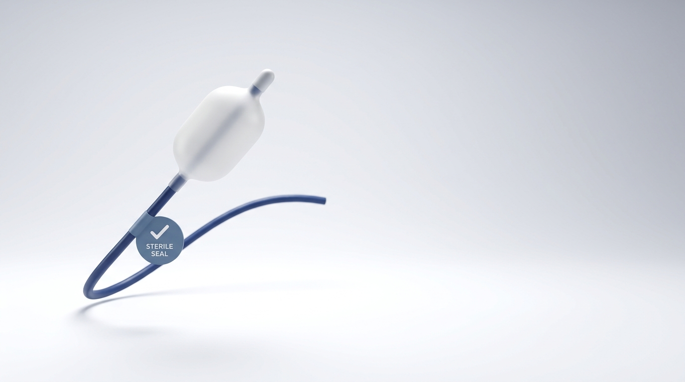
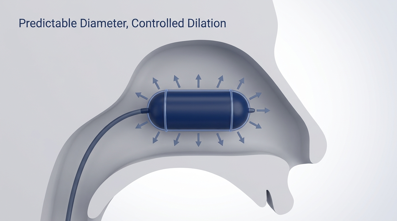
Balloon-assisted techniques aim to remodel selected deviations with less dissection in some cases, but they are generally not designed to shave down or remove discrete bony/cartilaginous spurs. If your main issue is a true spur, your ENT will typically recommend traditional instruments and techniques rather than a balloon-based approach.
If you’re new to this topic, start here: balloon-assisted septoplasty: https://www.clearpathnasal.com/blog/what-is-balloon-assisted-septoplasty.
Disclosure: ClearPath is a commercial device from our company. The discussion here is informational and not a product recommendation. Any decision about approach or device should be made with your treating ENT based on your anatomy and goals.
A practical next step is to ask your ENT whether you have a discrete spur that requires traditional correction and whether any less-invasive options are appropriate for your overall deviation pattern.
In short: balloon tools may help in select deviations, but true spurs typically need traditional spur-focused techniques.
Recovery and Results: What Improves After Spur/Septal Deformity Correction?
Symptom goals (what most patients care about)
- Easier nasal breathing (often most noticeable on the previously blocked side)
- Less nighttime mouth breathing in some people (snoring may or may not change and is not a reliable or primary expected result)
- Reduced irritation or nosebleeds in selected cases (remember that epistaxis has many causes beyond septal anatomy)
What can limit improvement (realistic expectations)
- Turbinate hypertrophy
- Nasal valve narrowing/collapse
- Allergies or chronic rhinitis
- Chronic sinusitis
Sometimes septoplasty is combined with turbinate reduction or sinus procedures when appropriate. (AAO-HNS, 2015; Iowa Protocols, 2023)
In short: septoplasty can address structural narrowing, but overall results depend on other nasal factors that may also need attention.
Questions to Ask Your ENT (Quick Checklist)
- Is my spur actually causing my symptoms?
- Is the spur anterior or posterior—and does that change the approach?
- Will you need to elevate one flap or both?
- How do you reduce the risk of mucosal tears and perforation?
- Are other causes of blockage present (turbinates, nasal valve, allergies)?
FAQs
Can a septal spur go away on its own?
A bony/cartilaginous spur typically does not “dissolve.” Symptoms can fluctuate (for example with allergies, swelling, or dryness), but the underlying structure generally remains.
If I have a spur, do I automatically need septoplasty?
No. A spur on imaging alone usually isn’t enough. Septoplasty for septal spur is most often considered when symptoms (especially obstruction) match the exam findings. (StatPearls; AAO-HNS 2015)
Is endoscopic septoplasty “safer” than traditional septoplasty?
Not proven overall. Endoscopic visualization can help surgeons perform more targeted work—particularly for posterior anatomy—but published data have not established that endoscopic septoplasty is safer across the board than traditional approaches. Risks still exist, and outcomes depend on anatomy, technique, and surgeon experience. (Kulkarni 2015)
What’s the biggest risk specific to spur work?
Mucosal tears are a key concern because the lining over a spur can be thin and adherent. Avoiding opposing tears on both sides in the same spot is an important part of reducing septal perforation risk. (StatPearls; Iowa Protocols, 2023)
Key Takeaways (Skimmable Summary)
- A septal spur alone doesn’t always require surgery—symptoms and exam guide candidacy.
- A mucoperichondrial/mucoperiosteal flap is the thin lining lifted to reach septal cartilage/bone safely.
- Bigger corrections may require wider or bilateral flap elevation.
- A posterior septal spur may be approached with endoscopic septoplasty and more targeted exposure, though local flap elevation is still required.
- Protecting the septal lining is central; tears—especially on both sides in the same spot—raise concern for perforation.
Citations
- StatPearls. Septoplasty. NCBI Bookshelf. https://www.ncbi.nlm.nih.gov/books/NBK567718
- University of Iowa Head & Neck Protocols (2023). Septoplasty for Nasal Obstruction: Indications and Techniques. https://www.iowaprotocols.medicine.uiowa.edu/protocols/septoplasty-nasal-obstruction-indications-and-techniques
- Kulkarni et al. (2015). Endoscopic Septoplasty: A Retrospective Analysis of 415 Cases. https://pmc.ncbi.nlm.nih.gov/articles/PMC4575672
- Han et al. (2015). Clinical Consensus Statement: Septoplasty. https://aao-hnsfjournals.onlinelibrary.wiley.com/doi/10.1177/0194599815606435
Disclaimer
This article is for general education and isn’t a substitute for personalized medical care. Please consult a qualified healthcare provider for diagnosis and treatment. An ENT evaluation is the best way to determine whether your spur is truly driving symptoms and what approach fits your anatomy.

Dr. David G. Dillard
Author
Share it with others!