What Is the Perpendicular Plate of the Ethmoid?
If you’ve been told you have a deviated septum (septal deviation) or you’re trying to understand your nasal anatomy before treatment, one structure that often comes up is the perpendicular plate of the ethmoid. It’s a small, thin bone—but because it sits right in the middle of the nose, it can play an outsized role in airflow and nasal obstruction.
A helpful way to picture it: think of the nasal septum as the interior dividing wall between two narrow hallways. Even if that wall is only slightly off-center, one hallway can start to feel cramped—especially when the lining swells from allergies or a cold.
Quick answer (for readers who want the definition fast)
What it is
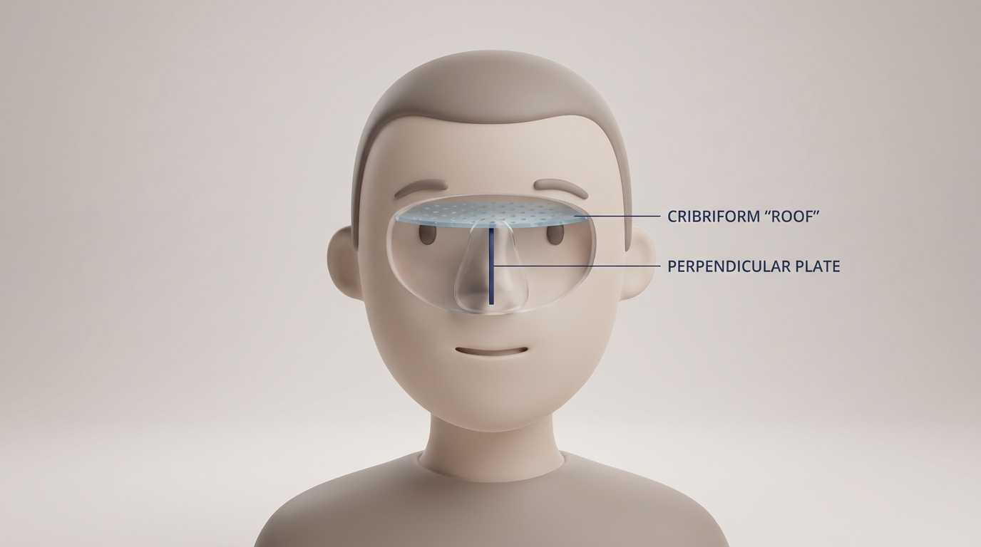
The perpendicular plate of the ethmoid is a thin, vertical piece of bone in the center of the nose. It extends downward from the cribriform plate (part of the ethmoid bone at the roof of the nasal cavity). (AnatomyZone; TeachMeAnatomy, 2025)
Why it matters
This bone helps form the nasal septum, the internal wall that separates the left and right nostrils. Because it’s thin and positioned along the midline, it’s commonly involved in deviations and injury, which can affect breathing. (Kenhub; TeachMeAnatomy, 2025)
*In short: a small midline bone that can have a big impact on airflow.*
Where is the perpendicular plate located?
The ethmoid bone’s “centerpiece” in the nasal cavity
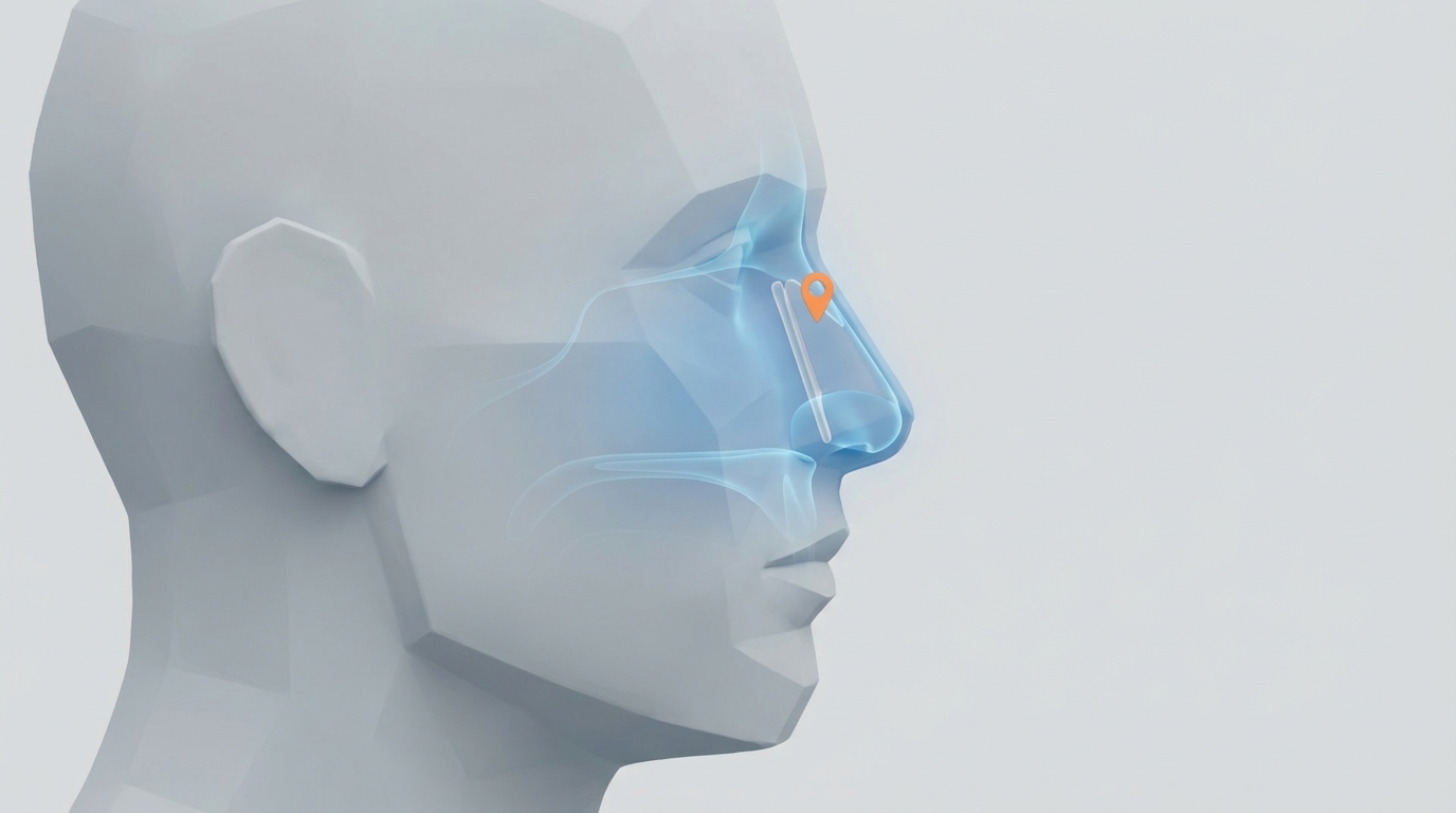
The ethmoid bone is a light, complex bone located between the eyes that contributes to the structure of the nasal cavity. One part of it forms the roof of the nasal cavity (the cribriform plate), and from there the perpendicular plate drops down like a vertical sheet. (TeachMeAnatomy, 2025; Kenhub)
In practical terms: the perpendicular plate is a midline bony divider sitting high inside the nose—right where a lot of airflow dynamics and tight spaces matter.
A quick “mental map” of where it sits
If you touch the outside bridge of your nose, you’re still far from the perpendicular plate. This structure is internal and higher up, behind the front cartilage many people picture when they think “septum.” That’s one reason high deviations can be hard to describe based on symptoms alone: the problem may feel “deep” or “up inside,” even though it’s still part of the septum.
What “upper bony portion of the septum” means
You may read that the perpendicular plate of the ethmoid makes up the “upper bony portion” of the nasal septum—the top segment inside the nose, behind the bridge area. (AnatomyZone; TeachMeAnatomy, 2025)
*Think of it as the upper, deeper bony segment of the internal dividing wall.*
What does it connect to (and what else makes up the septum)?
The 3 main septum components
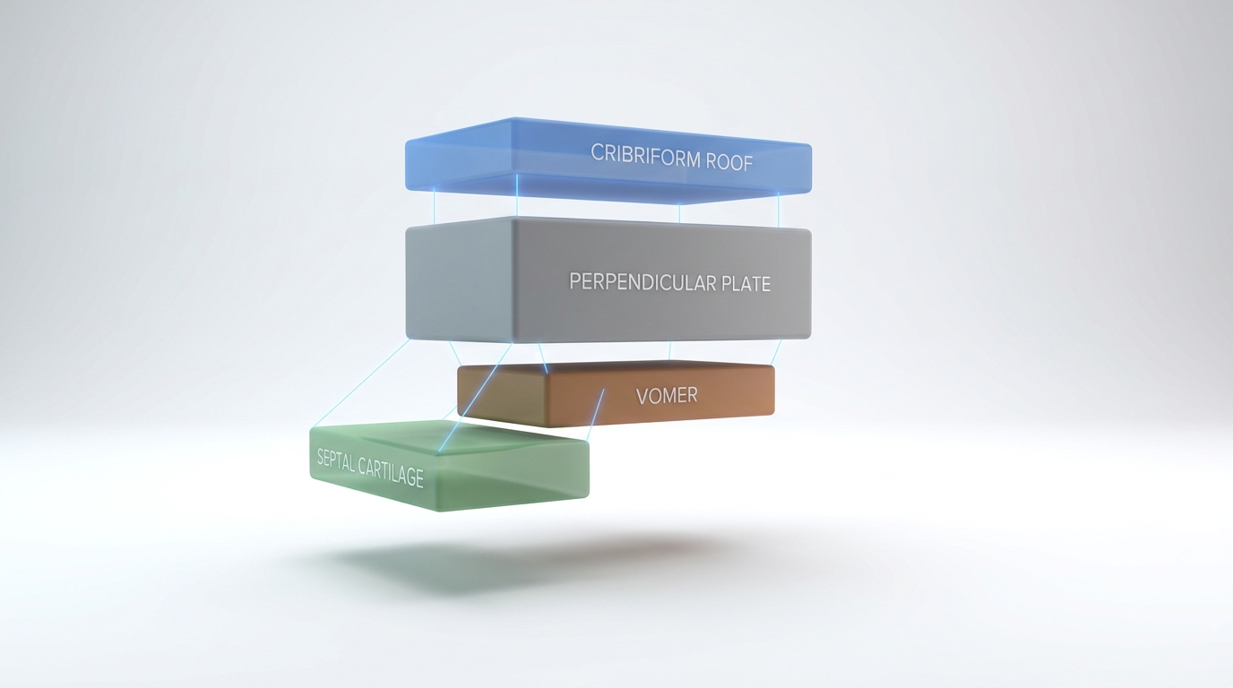
The nasal septum isn’t a single piece. It’s made from three main components:
- Perpendicular plate of ethmoid (upper bony part)
- Vomer (lower/posterior bony part)
- Septal cartilage (front, flexible part)
If you want a deeper, visual-friendly explanation of how these pieces fit together, see this overview of how the nasal septum is built: https://www.clearpathnasal.com/blog/deviated-septum-causes-understanding-your-nasal-anatomy
Key joints/articulations
The perpendicular plate helps complete the septum by connecting to other septal structures:
- It articulates inferiorly with the vomer
- And anteriorly with the septal cartilage (Kenhub; AnatomyZone)
These connections help explain why septal deviation varies from person to person: the septum can be off-center due to bone, cartilage, or both, and in different locations along its length.
*Bottom line: multiple parts form the septum, and the perpendicular plate links directly to both bone and cartilage.*

What does the perpendicular plate do?
Supports the nasal septum
The perpendicular plate provides structural support to the upper septum. Think of it as part of the internal framework that helps maintain the nose’s shape and keeps the left and right nasal passages separated.
Helps maintain nasal airflow efficiency
Airflow through the nose is affected by the size and shape of each nasal passage. When the septum is closer to midline, air often moves more freely. When the septum shifts to one side (septal deviation), one side may narrow, which can contribute to the feeling of blockage—especially during colds or allergy flares. Even small changes in nasal width can affect airflow and the sensation of obstruction.
A concrete example
If your “normal” airway is already a bit narrow on the left due to septal shape, then a routine cold can add swelling on top of that—turning a mild pinch point into a noticeable obstruction.
Plays a role in surgical planning
ENT clinicians assess where the septum is deviated (bony vs cartilaginous, front vs back, high vs low). Because the perpendicular plate is part of the upper bony septum, it’s one of the structures that may be evaluated when planning a correction.
*Functionally, the perpendicular plate is both a support strut and a potential pinch point for airflow.*
Common problems involving the perpendicular plate
Septal deviation (when the septum is off-center)
Septal deviation is common, and the perpendicular plate is often involved because it is thin and located on the midline. Even a modest shift can reduce space in one nasal passage and contribute to nasal obstruction. (TeachMeAnatomy, 2025; Kenhub)
It can also be “mixed”: a person may have some cartilage deviation in front and a higher bony deviation behind it. That’s why the exam often focuses on identifying all meaningful narrowing points, not just the most obvious one.
Nasal trauma
Nasal trauma—sports injuries, falls, or accidents—can change septal alignment. While many people think of the “nose bone” when they imagine a broken nose, internal structures can shift too. Because the perpendicular plate is a delicate bony sheet within the septum, it can be involved in septal injury, especially when combined with cartilage bending.
Why “thin bone” can still cause big symptoms
Nasal breathing is sensitive to small changes. Even subtle narrowing in the wrong spot can noticeably change airflow or create turbulence. That’s why a structure as thin as the perpendicular plate of the ethmoid can still be relevant when someone has persistent obstruction.
*Small structural shifts in a tight space can add up to big breathing changes.*
Symptoms that may be linked to septal deviation (including bony deviation)
Breathing and airflow symptoms
- One-sided nasal blockage or reduced airflow through one nostril
- Mouth breathing, especially at night
- Symptoms that feel worse during colds/allergies (when lining swelling makes a narrowed passage even tighter)
Quality-of-life symptoms some patients report
- Sleep disruption or snoring
- Feeling “stuffy” much of the time
- Facial pressure or headache (often multifactorial and not specific to septal deviation alone)
When symptoms suggest it’s time for an ENT evaluation
- Persistent nasal obstruction despite appropriate medical therapy
- Recurrent nosebleeds
- Frequent sinus infections
- A history of nasal trauma
- Major sleep impact
*If symptoms persist or escalate, a focused exam can clarify whether structure, swelling, or both are driving the blockage.*

How ENT clinicians evaluate septal anatomy (including the perpendicular plate)
History + nasal exam
Clinicians usually start with questions about:
- Which side feels blocked (or if it alternates)
- Allergy symptoms, sinus history, and medication trials
- Prior injuries or surgeries
- Sleep and exercise limitations
Then they examine the nasal cavity to look for deviation, swelling, and other contributors.
Nasal endoscopy (when needed)
A nasal endoscopy uses a thin camera to view deeper inside the nose. It can help clarify what’s causing obstruction—especially when the deviation is higher or more posterior, or when swelling obscures the view.
In practical terms, endoscopy can help answer questions like: is the narrowing mostly from septal shape, from swollen tissues, or from a combination?
CT imaging (only in specific situations)
CT scans are typically reserved for situations like suspected sinus disease, complex anatomy, or detailed surgical planning. They’re not always required for a straightforward deviated septum workup.
*Evaluation aims to map symptoms to anatomy so treatment can be tailored appropriately.*

Treatment options if the septum is deviated
Non-surgical symptom management
Some people improve with measures aimed at reducing swelling and irritation, such as:
- Saline rinses
- Allergy management
- Nasal steroid sprays (when appropriate)
- Addressing turbinate swelling
If you’re exploring this route first, this guide may help: https://www.clearpathnasal.com/blog/can-you-fix-a-deviated-septum-without-surgery
*For many, reducing lining swelling can lessen symptoms even when anatomy is not perfectly midline.*
Septoplasty (structural correction)
Septoplasty is a procedure designed to straighten the septum to improve airflow. Depending on your anatomy, septoplasty may address:
- Cartilage deviation (front portion)
- Bony deviation (including the vomer and/or parts associated with the ethmoid region)
If you’re comparing options and recovery expectations, here’s a side-by-side overview: https://www.clearpathnasal.com/blog/clearpath-vs-traditional-septoplasty-recovery-safety-results
*The goal is functional: to create a more balanced, open nasal airway.*
Minimally invasive approaches (where appropriate)
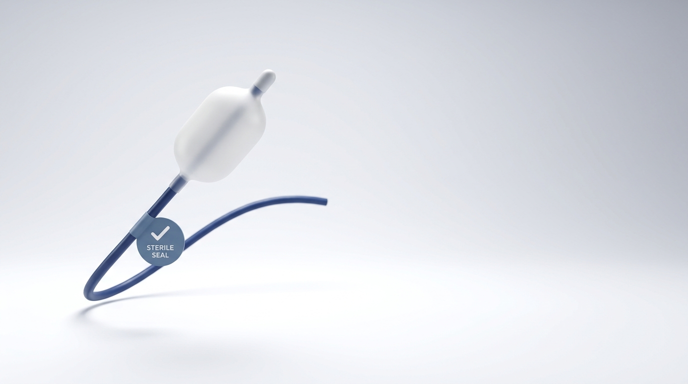
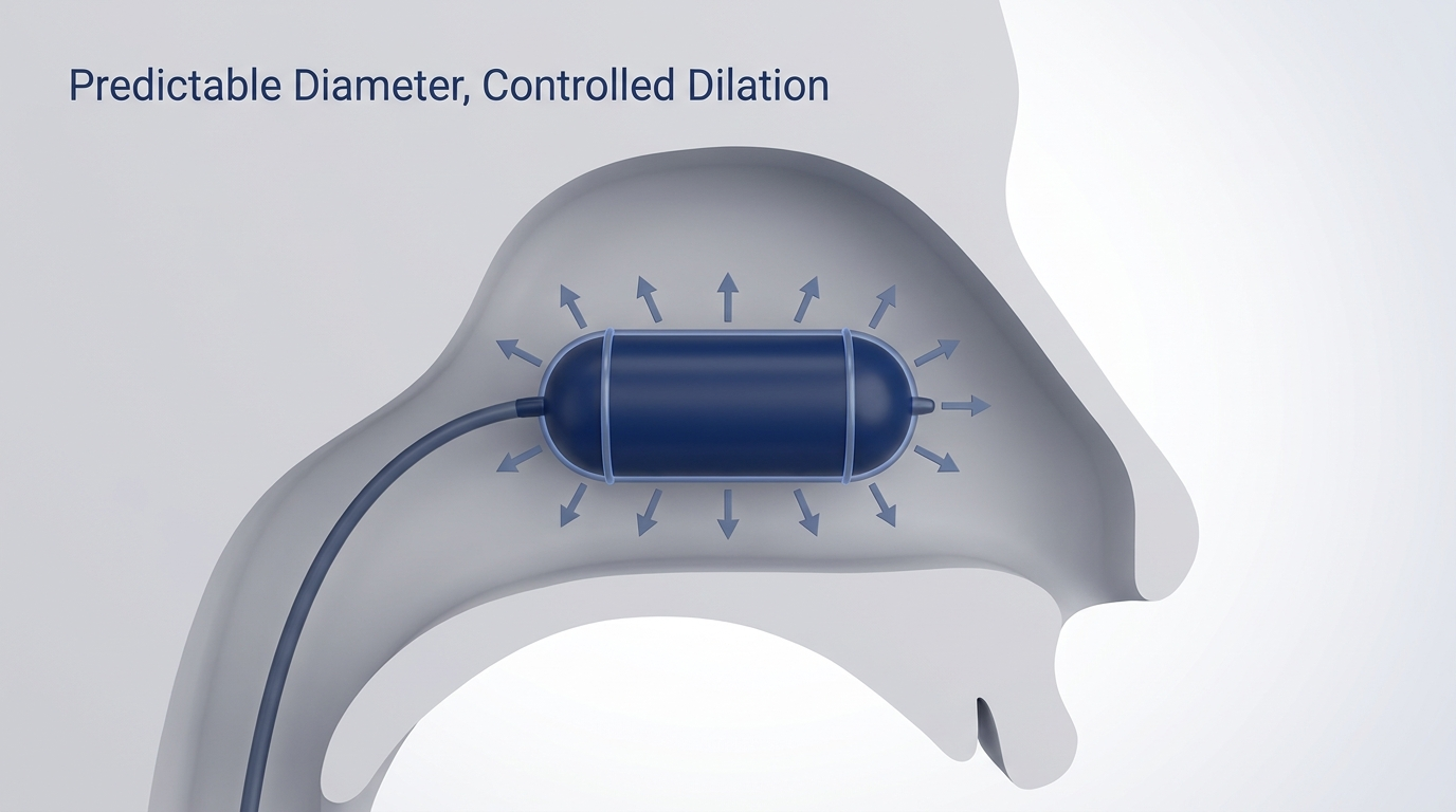
For selected anatomy types, some ENT surgeons may use minimally invasive or catheter-based, balloon-assisted techniques to help improve septal alignment while aiming to limit tissue disruption. Availability and suitability depend on the surgeon’s assessment and your individual anatomy. To learn the basics, see this overview of balloon septoplasty: https://www.clearpathnasal.com/blog/what-is-balloon-septoplasty-a-minimally-invasive-option-for-deviated-septum
What does research show? (One example)
In one retrospective review of 107 consecutive CNB-assisted septoplasty cases, the authors reported improvement in alignment measurements and patient-reported outcomes, with no major complications in that cohort. In a subgroup of 23 patients with pre- and post-operative CT, septal alignment symmetry improved 22% at the nasolacrimal duct and 45% at the point of maximal deviation, and SNOT-22 scores improved from 58.7 to 44.8 at one month (a 23.8% reduction). These findings reflect a single-center retrospective experience and may not generalize to all patients or techniques. (Dillard et al., 2026)
*Evidence continues to evolve; discuss benefits and risks with your clinician in the context of your anatomy and goals.*
FAQs (patient-friendly)
Is the perpendicular plate the same thing as the nasal septum?
No. The perpendicular plate of the ethmoid is one bony part of the septum. The septum also includes the vomer and septal cartilage. (Kenhub; AnatomyZone)
Can a deviated septum get worse over time?
Sometimes symptoms become more noticeable over time due to swelling patterns, additional minor trauma, or aging changes in nasal tissues—even if the underlying anatomy is similar. Read more on whether a deviated septum can get worse over time: https://www.clearpathnasal.com/blog/does-a-deviated-septum-get-worse-over-time
Can you feel the perpendicular plate from inside your nose?
Not directly. People typically feel obstruction due to the overall septum shape plus nasal lining swelling, not because they can sense a specific bone.
Do all septal deviations need surgery?
No. If symptoms are mild or improve with medical management, surgery may not be necessary. When obstruction is persistent and clearly structural, procedures like septoplasty (including, in selected cases, minimally invasive or balloon-assisted approaches) may be discussed with an ENT.
*Most people start with medical therapy; structural procedures are considered when symptoms remain clearly anatomy-driven.*
Key takeaways
- The perpendicular plate of the ethmoid is a thin vertical bone descending from the cribriform plate that forms the upper bony portion of the nasal septum. (AnatomyZone; TeachMeAnatomy, 2025)
- It connects with the vomer and septal cartilage, completing the bony/cartilaginous septum. (Kenhub; AnatomyZone)
- Because it’s thin and midline, it’s commonly involved in septal deviation and nasal trauma, which can contribute to nasal obstruction and may prompt consideration of septoplasty in selected patients. (TeachMeAnatomy, 2025; Kenhub)
*Understanding where and how the septum deviates helps match symptoms to the most appropriate treatment plan.*
Next step if symptoms persist
If nasal blockage is persistent or affecting sleep, consider scheduling a visit with an ENT to review the cause of your symptoms and discuss appropriate treatment options for your anatomy.
References
- AnatomyZone. Perpendicular Plate (Ethmoid Bone). https://anatomyzone.com/articles/perpendicular-plate-ethmoid-bone/
- TeachMeAnatomy (2025). The Ethmoid Bone. https://teachmeanatomy.info/head/osteology/ethmoid-bone/
- Kenhub. The Ethmoid Bone. https://www.kenhub.com/en/library/anatomy/the-ethmoid-bone
- Wikipedia. Perpendicular plate of ethmoid bone. https://en.wikipedia.org/wiki/Perpendicular_plate_of_ethmoid_bone
- Dillard J, Koudouovoh C, Lee V, et al. Rhinology Online. 2026;9:18–23. doi:10.4193/RHINOL/25.018
This article is for educational purposes only and is not medical advice. Please consult a qualified healthcare provider for diagnosis and treatment.

Dr. David G. Dillard
Author
Share it with others!